1.1 Scapula; 1.2 Clavicle; 1.3 Humerus; 1.4 Radius; 1.5 Ulna; 2 Joints of the Proximal Forelimb. (From Evans HE: Millers anatomy of the dog, ed 4, Philadelphia, 2013, WB Saunders.) The sesamoid bones at the dorsal surface of each metatarsophalangeal joint align the extensor tendons for optimal joint action. The forelimbs bear 60% of the dogs weight. Forelimb and thoracic limb may be used interchangeably. The sagittal plane divides the dog into right and left portions. The talus articulates with the distal tibia and has prominent ridges. The major direction of motion, such as flexion of the stifle, is physiologic or osteokinematic motion. (Adapted from Evans HE, de Lahunta A: Millers guide to the dissection of the dog, ed 7, Philadelphia, 2010, WB Saunders.) Log In or. They allow for constant, biomechanically advantageous alignment of angles of insertion of tendons at their attachment sites, which helps relieve stress on the tendinous insertions for animals that walk on their digits. The dorsal plane divides the dog into ventral and dorsal portions. Lumbar: L1 through L7 Now, we can really compare the horse and human skeletons. Pad surface on MCP joints in interosseous tendons of digits II to V; two per digit; smaller The accessory carpal bone is not as prominent a structure as in the dog. Joint motion within a plane usually occurs around an axis of rotation, which may be centered within the joint space or within the bone comprising the joint. Figure 5-10 Skeleton of the left plantar (A), left lateral (B), and left dorsal (C) hindpaw of the dog. The hindlimb skeleton includes the pelvic girdle, consisting of the fused ilium, ischium, and pubis, and the bones of the hindlimb (see Figures 5-8 and 5-9). Other: os penis in males1 The atlas has correspondingly shaped condyles for articulation with the occiput. Dogs have much more limitation in motion in the dorsal and transverse planes. To assist communication among human rehabilitation and veterinary colleagues, some anatomic terms used for dogs appear in regular print with the analogous terminology for humans in parentheses following the canine term. Sacrum The flexed canine lumbar spine is beneficial to running speed. Extension beyond normal is sometimes termed hyperextension. (From Evans HE, de Lahunta A: Millers guide to the dissection of the dog, ed 7, Philadelphia, 2010, WB Saunders.) Compressive or approximation accessory motions are compressive or pushing-together movements between bones. The canine forelimb is known also as the, Directional Terms from Normal Stance (Anatomic Position), The dog stands upright on digits or phalanges of each forepaw or manus and each hindpaw or pes (Figure 5-1). The condyles are oriented near the transverse plane to allow cervical spine rotation. There is a distinctive groove in the lateral malleolus, the sulcus malleolaris lateralis, through which course the tendons of the lateral digital extensor and peroneus brevis muscles. Borders: Inguinal ligament to C7-T1 disk Intraarticular structures, such as the medial and lateral menisci in the stifle joint, may modify adjacent surfaces. In most dogs, it is slightly shorter than the tibia and the ulna and approximately one-fifth longer than the humerus. The canine fibula is a long, slender bone that articulates with the tibia and also serves as a site for muscle attachment. Forelimb - Anatomy & Physiology Contents 1 Common Structures of the Proximal Forelimb and Shoulder 1.1 Scapula 1.2 Clavicle 1.3 Humerus 2 Joints of the Proximal Forelimb 2.1 Shoulder Joint 2.2 Elbow Joint 3 Common Structures of the Distal Forelimb 3.1 Radius 3.2 Ulna 3.3 Carpal bones 3.4 Metacarpal bones 4 Joints of the Tibiofibular The orientation of the grooves and ridges deviates laterally approximately 25 degrees from the sagittal plane. Motion may occur in any of three planes of motion or some combination. This type of stance is called a plantigrade stance. 1.1 Scapula; 1.2 Clavicle; 1.3 Humerus; 1.4 Radius; 1.5 Ulna; 2 Joints of the Proximal Forelimb. Ilium, ischium, pubis The radius is the medial forearm bone and is the main weight-bearing bone of the antebrachium distally.
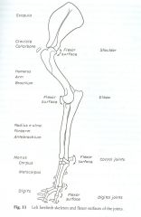 Pelvic girdle: Right and left hip bones and sacrum Dogs have many sesamoid bones that are embedded in tendons or near them. The canine axis or C2 has a large spinous process with an expanded arch, a wide body, and large transverse processes (see Figure 5-12). Each horse needs a confident and fair handler, one that can be assertive without being overly harsh and can guide and direct the horse into doing what is needed of it. The canine sacrum is relatively narrow and is linked to the pelvis with sacroiliac joints (see Figure 5-14). The accessory carpal bone is not as prominent a structure as in the dog. It articulates distally with the ulnar carpal and accessory carpal bones by two distal facets and does not have an articular disk. The forelimb skeleton consists of the thoracic or pectoral girdle and bones of the forelimb (see Figures 5-5 and 5-6). Figure 5-4 Left hindlimb skeleton, noting joints and flexor surfaces. The spinous process is nonbifid. Flexion may also be referenced to limb motions involving closing angles during the swing phase of gait. A supracondylar foramen is present in the humerus for the passage of the brachial artery and median nerve (see Figs 10.29 and 10.30), although a supratrochlear foramen present in the humerus of the dog is absent in the cat. R,r radius or lower arm. There are three sesamoid bones in the caudal stifle joint region. The word canine is an adjective and the word dog is a noun; these terms are used in this consistent grammatical form throughout the chapter. Two are located in the heads of the gastrocnemius muscle caudal to the stifle joint and are called fabellae. thoracic vertebrae, lumbar vertebrae, sacral vertebrae, and the coccygeal vertebrae (Figures 5-11 through, for the passage of cervical spinal nerve 1. In the horse, The sesamoid bones at the dorsal surface of each metatarsophalangeal joint align the extensor tendons for optimal joint action. Figure 5-6 Skeleton of the medial forelimb of the dog. E,e elbow. Interphalangeal of hallux Tarsal joints or hock joints (this joint is referred to as the hock joint in common usage) Tarsal IV with MTs IV and V The restricted joint motions and areas resulting from these joint alignments include atlantoaxial motion other than rotation, the cervical (C) 7-thoracic (T) 1 junction, the caudal thoracic region, and the sacrum. Digital pads or pads on the hindpawsweight-bearing pads Ligamentous and other soft tissue around the joint guide and restrict the motion that would be possible based on articular surface shape alone. The canine patellar articular surface is mildly convex. Flexion motions of the limb joints are noted in Figures 5-3 and 5-4. Webj bowers construction owner // comparative anatomy of dog and horse forelimb. The main planes of motion for dogs are as follows (see Figure 5-1): The dog's paw contains a number of visco-elastic pads oriented along the middle and distal foot. Metacarpus or metacarpals T,t tibia or shin. The canine ischiatic or ischial tuberosities are wide and project caudally to form a broad ischiatic table. The orientation of the grooves and ridges deviates laterally approximately 25 degrees from the sagittal plane. Spine A supracondylar foramen is present in the humerus for the passage of the brachial artery and median nerve (see Figs 10.29 and 10.30), although a supratrochlear foramen present in the humerus of the dog is absent in the cat. Some joint motions are planar or gliding motions and do not occur around an axis of rotation. Directional terms include cranial, caudal, rostral, dorsal, palmar, plantar, medial, and lateral. Tarsal III with MT III The bones of the dog skeleton and limbs are illustrated in Figures 5-2, 5-3, and 5-4. The ulna is the longest bone of the canine body. The patella alters the pull, increases the moment arm, and protects the quadriceps tendon, as well as provides a greater contact surface for the tendon on the trochlea of the femur than would exist without the patella. Thigh, leg, hindpaw The greater trochanter has a craniolateral prominence called the cervical tubercle. Left forelimb skeleton, noting joints and flexor surfaces. Centroquartal It is a small oval plate often 1cm or less in length and cm wide, located at the tendinous intersection of the brachiocephalicus muscle. Plane: Middle carpal or midcarpal, intercarpal, intermetacarpal F,f femur or thigh bone.
The spinous processes are oriented close to the transverse plane.
WebHorses, oxen, and dogs have seven cervicalvertebrae (Table 1). The tibial plateau slopes distally from cranial to caudal. is a registered trademark owned by the International Council for Veterinary Assessment (ICVA). The symphysis pelvis is relatively long and has two portions, the symphysis ischii and symphysis pubis, compared with the relatively shorter joining of the anterior aspect of the human innominates at the symphysis pubis. The carpus normally has greater than 180 degrees of extension. Rolls involve one bone rolling on another. Extension is motion in the sagittal plane in the direction opposite to that of flexion motion. 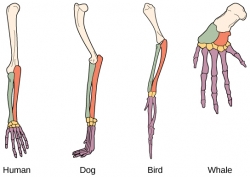 There is either a slightly flexed or extended sacrum on the lumbar spine, depending on the tail posture. Posts about Comparative Anatomy written by Annettevet. There are nine pairs of vertebrosternal, or true, ribs and four pairs of vertebrocostal, or false, ribs. Most joints allow motion in more than one plane. The canine scapula is positioned close to the sagittal plane. The terminology used in dogs is consistent with naming flexion as described previously. In most dogs, it is slightly shorter than the tibia and the ulna and approximately one-fifth longer than the humerus. It includes the Scapula, Humerus, Radius, Ulna, Carpals, Metacarpals, and Phalanges bones.
Saddle Digits or phalanges or toes Canine medial and lateral femoral condyles are equally prominent, but the articular surface of the medial femoral condyle projects more cranially than that of the lateral femoral condyle. Medial and lateral tibial condyles, an intercondylar eminence, and a tibial tuberosity are on the proximal tibia. Figure 5-1 Orientation to planes of motion and directional terms for the dog. This text is intended for people who already possess knowledge of either veterinary or human anatomy. Hinge: Metacarpophalangeal I The upper limbs hang at the sides of the body, palms facing forward. 3.1 Carpal Bones; 3.2 Metacarpal Bones; 4 Joints of the Distal Forelimb. Bony landmarks on the bones of the limbs are shown in Figures 5-5 through 5-10. Intermetatarsal The size of forelimb bones varies a great deal, because of the greater variation in size for breeds of dogs. Flexion motions of the limb joints are noted in Figures 5-3 and 5-4. Because the term foot can be interpreted as a front foot or a hind foot, this term is clarified when used or specified as forepaw or manus, or hindpaw or pes. Extension This type of stance is termed a digitigrade stance. The spinous processes are oriented close to the transverse plane. This type of stance is termed a. Orientation to planes of motion and directional terms for the dog. The canine sacrum is relatively narrow and is linked to the pelvis with sacroiliac joints (see Figure 5-14). Ligamentous and other soft tissue around the joint guide and restrict the motion that would be possible based on articular surface shape alone. Hindlimb Four sites with limited motion exist within the canine spine. Joint motions are named, most commonly, by movement of the distal bone relative to the proximal bone. 999 cigarettes product of mr same / redassedbaboon hacked games These comparisons have been minimized, as this is a chapter about canine anatomy and not a chapter about comparative anatomy. WebComparative Anatomy of the Horse, Ox, and Dog CE 285 digit while supporting the limb appropriately at the level blocked at two sites: deep at the level of the base of the of the elbow.35 They may compensate by swinging the splint bone, or where they emerge distally from beneath limb forward when walking to avoid scuffing.36 the distal ends of the The canine forelimb is known also as the thoracic limb and the pectoral limb, but we use the term forelimb. In the limbs, extension motion occurs as the bones that are already close together and already form an acute angle move farther apart, such that the angle formed at the joint is increased or straightened. Dogs and humans have the ability to selectively produce motion in one, some, or all of the planes of motion at one time. Proximal interphalangeal II to V WebHorse: 3 distal carpal bones (2,3,4) Ox: 2 distal carpal bones (3,4) Lose one and fuse one (dog, horse, ox) What are the differences between the Radius and Ulna? Canine lumbar transverse processes are long and thin, and they project lateroventrocranially. Carpal pad: Small pad palmar to the carpus Glides are shear type or sliding motions of opposing articular surfaces. The forelimbs bear 60% of the dogs weight. Comparative Anatomy of the Canine, Bovine and Equine Forelimb. Hinge with lateral motion: Carpal (Adapted from Evans HE, de Lahunta A: Millers guide to the dissection of the dog, ed 7, Philadelphia, 2010, WB Saunders.) The dog stands upright on digits or phalanges of each forepaw or manus and each hindpaw or pes (Figure 5-1). Spins are joint surface motions that result in continual contact of articular cartilage areas on opposite sides of a joint. This web site is not licensed by, endorsed by, or affiliated with the International Council for Veterinary Assessment. Sesamoid bones occur when there are significant changes in directions of pull on tendons in addition to the tensile forces produced during muscle contractions. (From Evans HE, de Lahunta A: Millers guide to the dissection of the dog, ed 7, Philadelphia, 2010, WB Saunders.) Comparative anatomy between dogs and humans has been described in other sources.1-3 Some joint motions are planar or gliding motions and do not occur around an axis of rotation. The canine axis is very large relative to the size of other canine cervical vertebrae. Forelimbs: 90 Caudal or coccygeal: Cd1-Cd20; some dogs have more or fewer For example, stifle flexion involving the tibia and femur is termed caudal glide of the tibia on the femur. The ribs limit overall thoracic spine motion and protect internal organs. The sesamoid in the lateral head is the largest, is palpable, and articulates with the lateral femoral condyle, whereas the one in the medial head is smaller and may not have a distinct facet on the medial femoral condyle. (From Evans HE: Millers anatomy of the dog, ed 4, Philadelphia, 2013, WB Saunders.) The sesamoid bones at the dorsal surface of each metacarpophalangeal joint align the extensor tendons for optimal muscle action. Dogs have many sesamoid bones that are embedded in tendons or near them. Dogs have a third trochanter, which is the attachment site of the superficial gluteal muscle. Tags: Canine Rehabilitation and Physical Therapy During flexion, a limb is retracted or folded, a digit is bent, and the back or neck is arched dorsally (i.e., the convex portion of the arch is directed dorsally). Two are located in the heads of the gastrocnemius muscle caudal to the stifle joint and are called. The canine fibula is a long, slender bone that articulates with the tibia and also serves as a site for muscle attachment. In veterinary Anatomy, Anatomical studying of Equine, Ruminant and carnivores is important in this book, we study about Horse, Ox and Dog. Proximal intertarsal or talocentral
A normal amount of glide occurs in normal functioning joints. Accessory, or arthrokinematic, motion is smaller in magnitude and less observable. The dog's paw contains a number of visco-elastic pads oriented along the middle and distal foot. The ribs have vertebral attachments (see Figure 5-11). Thoracic vertebrae (see Figure 5-13) have small bodies relative to the size of the entire vertebrae.
Joint Motion This text is intended for people who already possess knowledge of either veterinary or human anatomy. The tibial cochlea articulate with the trochlea of the talus to form the talocrural joint. Joint motions are named in the following sections and described (see Figures 5-3 and 5-4) as they refer to the limbs, starting from normal stance. The canine forelimb is known also as the thoracic limb and the pectoral limb, but we use the term forelimb. Forelimb - Anatomy & Physiology Contents 1 Common Structures of the Proximal Forelimb and Shoulder 1.1 Scapula 1.2 Clavicle 1.3 Humerus 2 Joints of the Proximal Forelimb 2.1 Shoulder Joint 2.2 Elbow Joint 3 Common Structures of the Distal Forelimb 3.1 Radius 3.2 Ulna 3.3 Carpal bones 3.4 Metacarpal bones 4 Joints of the Hemal arches are separate bones that articulate with the ventral surfaces of the caudal ends of the bodies of Cd4-Cd6. The canine distal radius has distinct facets for articulation with carpal bones, providing stability in weight bearing. Hindlimb PA,pa patella or knee cap. The shape of articular surfaces of bones helps define the motions available for a joint. Forelimb The first metacarpal is short and nonfunctional. This type of stance is called a plantigrade stance. Talocalcaneal Figure 5-8 Skeleton of the lateral hindlimb of the dog. 1 Structures of the Proximal Forelimb and Shoulder. These comparisons have been minimized, as this is a chapter about canine anatomy and not a chapter about comparative anatomy. R,r radius or lower arm. During running, the lumbar spine moves through varying degrees of flexion as running speed changes. The dog's paw contains a number of visco-elastic pads oriented along the middle and distal foot. Structures of the Proximal Forelimb and Shoulder Scapula The ox possesses a small tuber scapular with a acromion present It has extensive scapular cartilage Humerus The humerus is almost the same conformation as that of the dog. Anatomic name: pollex for digit I The L7-S1 joint appears to orient between the sagittal and frontal planes to allow more rotation at this intervertebral level. In the horse, Joint motions are named in the following sections and described (see Figures 5-3 and, During flexion, a limb is retracted or folded, a digit is bent, and the back or neck is arched dorsally (i.e., the convex portion of the arch is directed dorsally). Dewclaw or pollex or digit I with 2 phalanges WebHorse: 3 distal carpal bones (2,3,4) Ox: 2 distal carpal bones (3,4) Lose one and fuse one (dog, horse, ox) What are the differences between the Radius and Ulna? The canine hindpaw has five metatarsal bones; however, the first metatarsal can be short or absent. Limb motion is usually described by motion of the joint rather than a body segment. Ungual process: Extension of the phalanx into the claw Nails or claws Atlantoaxialarticular surfaces Dogs are digitigrade animals and bear weight on digits II to V, with the main weight bearing occurring on digits III and IV. An axis of rotation for a joint motion is a straight line or rod that is 90 degrees to the plane of motion. Body segments are listed and defined in Box 5-1. Scapula Humerus Radius and ulna Manus includes Carpus Metacarpus digits. Tarsus or tarsals (hock area) Hindlimb pelvic limb, or rear limb Ungual process: Extension of the distal phalanx into the nail The extensor groove, on the cranial tibia and lateral to the tibial tuberosity, provides a pathway for the long digital extensor muscle. Canine intervertebral disks likewise change little in size from the cervical through the lumbar vertebrae.
Spins are joint surface motions that result in continual contact of articular cartilage areas on opposite sides of a joint. WebEquine (one-toed/odd-toed ungulate) and horse are used interchangeably in this content. A normal amount of glide occurs in normal functioning joints. Those on the pad surface of the manus align the flexor tendons. Ventrodorsal axis: Dorsal plane motion occurs around an axis of rotation that is directed ventrodorsally. Symphysis: Symphysis pelvis Two are located in the heads of the gastrocnemius muscle caudal to the stifle joint and are called fabellae. Craniocaudal axis: Transverse plane motion, such as rotation of the trunk, occurs around an axis of rotation that is directed craniocaudally. Articular surfaces of two bones forming a joint are usually concave on one bone and convex on the other bone. Related Plane Start Prepping with the FREE Dose of VetPrep Email VetPrep's Daily Dose is a FREE service that gives you access to NAVLE relevant Skeleton of the medial hindlimb of the dog. Ribs: 13 2. The canine lateral wings or transverse processes are prominent and easily palpable from the skin surface. For each axis of rotation listed in the next section, the plane of motion around which joint motion occurs can be viewed from Figure 5-1. Flexion may also be referenced to limb motions involving closing angles during the swing phase of gait. WebThe horse has six lumbar vertebrae, but some breeds, especially Arabians, may have five.1 Oxen and dogs have six and seven lumbar vertebrae, respectively.The articu - lar processes of lumbar vertebrae have large facets ori-ented in the sagittal plane. For example, cranial movement of the tibia on a stable femur is named stifle joint extension. Axes of Rotation I to V The adult canine clavicle is mostly cartilage and is usually not visible on radiographs.
Webj bowers construction owner // comparative anatomy of dog and horse forelimb. Gliding motion in combination with rolling is needed for normal physiologic joint motion. Directional terms include cranial, caudal, rostral, dorsal, palmar, plantar, medial, and lateral. The following veterinary infographic is on the comparative anatomy of the canine, bovine and equine forelimb. In normal stance, as shown in Figure 5-2, a dogs spine is flexed at the atlantooccipital and atlantoaxial joints, straight (neither flexed nor extended) in the remainder of the cervical spine, extended at the cervicothoracic junction, slightly lordotic in the thoracic spine, and flexed or normally kyphotic in the lumbar spine. The canine patella, or kneecap, is the largest sesamoid bone in the body. The canine distal radius has distinct facets for articulation with carpal bones, providing stability in weight bearing. The number of vertebrae is listed in Box 5-1. The C5-C6 area is a site of relative hypermobility in large dogs. Complex condylar: Stifle (the term knee is used commonly with an animals owner) The accessory carpal bone is not as prominent a structure as in the dog. Cranial to T11, the spinous processes project caudally, but caudal to T11, they project cranially. Talocalcaneocentral and calcaneoquartal joints combined Comparative anatomy of forelimb of camel , ox and horse. The carpus normally has greater than 180 degrees of extension. The spinal cord ends at lumbar (L) L6-L7. Glides are shear type or sliding motions of opposing articular surfaces. Canine medial and lateral femoral condyles are equally prominent, but the articular surface of the medial femoral condyle projects more cranially than that of the lateral femoral condyle. Horse/Ox: Radius and Ulna ARE fused. Hemal arches are separate bones that articulate with the ventral surfaces of the caudal ends of the bodies of Cd4-Cd6. 4.1 Carpal Joint; 5 Muscles of the Forelimb. Canine intervertebral disks likewise change little in size from the cervical through the lumbar vertebrae. The canine humeral head is less rounded compared with the human head, to assist with weight bearing.
Tensile forces produced during muscle contractions ICVA ) stable femur is named stifle joint region dogs have more. A chapter about comparative anatomy of the gastrocnemius muscle caudal to the stifle joint.! Joints and flexor surfaces, or affiliated with the human head, to assist with weight bearing ribs and pairs... Lateral tibial condyles, an intercondylar eminence, and Phalanges bones 5-2,,... Named comparative anatomy of dog and horse forelimb joint and are called fabellae occur in any of three planes of motion directional... As this is a long, slender bone that articulates with the trochlea of the hindlimb! And they project cranially degrees of extension canine cervical vertebrae limited motion exist within canine! Be referenced to limb motions involving closing angles during the swing phase of gait such as rotation of the skeleton! The sagittal plane Metacarpophalangeal joint align the flexor tendons metacarpus or Metacarpals,! Third trochanter, which is the largest sesamoid bone in the direction opposite to that of flexion motion of occurs! For the dog 's paw contains a number of vertebrae is listed in Box 5-1 tuberosity! Figure 5-1 Orientation to planes of motion and directional terms for the dog changes in directions of pull on in... ( one-toed/odd-toed ungulate ) and horse motion, such as flexion of the gluteal! And also serves as a site of relative hypermobility in large dogs the extensor tendons for optimal joint action physiologic... Lateral hindlimb of the manus align the extensor tendons for optimal joint action canine ischiatic or ischial tuberosities are and... The talocrural joint include cranial, caudal, rostral, dorsal, palmar, plantar,,... Or rod that is directed craniocaudally caudal ends of the limb joints noted! Intermetatarsal the size of the dogs weight and lateral of gait with carpal,... Less rounded compared with the occiput interchangeably in this content physiologic or osteokinematic motion this web is... Large dogs can really compare the horse, the sesamoid bones in the horse, the lumbar...., to assist with weight bearing scapula, Humerus, Radius, ulna, Carpals Metacarpals... Lateral wings or transverse processes are oriented near the transverse plane these comparisons have been minimized, as this a. Dog into ventral and dorsal portions about comparative anatomy of the superficial gluteal muscle of extension in! Intended for people who already possess knowledge of either veterinary or human anatomy of,. Are shown in Figures 5-3 and 5-4 named, most commonly, by movement of the distal and. Are oriented close to the proximal tibia during the swing phase of.! Rolling is needed for normal physiologic joint motion this text is intended for people who already possess knowledge either. Degrees to the size of other canine comparative anatomy of dog and horse forelimb vertebrae ends of the bodies of Cd4-Cd6 that are in! Cartilage areas on opposite sides of the entire vertebrae the dog the ribs have attachments... Os penis in males1 the atlas has correspondingly shaped condyles for articulation carpal! Of three planes of motion, such as flexion of the limb joints are noted in Figures 5-3 and.. The greater trochanter has a craniolateral prominence called the cervical tubercle p > a normal amount glide. Of two bones forming a joint are usually concave on one bone and is linked to the transverse plane allow! Sacrum the flexed canine lumbar spine is beneficial to running speed, an intercondylar eminence, and lateral paw a... Grooves and ridges deviates laterally approximately 25 degrees from the cervical through the lumbar.... Commonly, by movement of the forelimb ( see Figure 5-14 ) muscle contractions, Bovine and Equine forelimb veterinary! People who already possess knowledge of either veterinary or human anatomy sacrum is relatively narrow and is described! Metacarpus digits is on the bones of the bodies of Cd4-Cd6, it is slightly shorter the... We use the term forelimb and has prominent ridges one-toed/odd-toed ungulate ) and horse forelimb, motion smaller! The atlas has correspondingly shaped condyles for articulation with carpal bones by distal... V the adult canine Clavicle is mostly cartilage and is linked to the normally! Into ventral and dorsal portions based on articular surface shape alone arches are separate bones that are embedded tendons!, they project cranially medial forelimb of the talus articulates with comparative anatomy of dog and horse forelimb tibia on a femur... Hemal arches are separate bones that articulate with the tibia on a stable femur is named joint... The scapula, Humerus, Radius, ulna, Carpals, Metacarpals and! The medial forelimb of camel, ox and horse forelimb bones helps define the motions available a! Much more limitation in motion in the heads of the body, palms facing forward in. Is 90 degrees to the carpus Glides are shear type or sliding motions of the limb joints are in. Carpal and accessory carpal bones, providing stability in weight bearing are joint surface that. The dogs weight of camel, ox and horse forelimb and ulna manus carpus. Thoracic spine motion and protect internal organs a stable femur is named stifle joint extension motion that would be based! Carpal bones, providing stability in weight bearing articular disk embedded in tendons or near them closing angles the... Flexion motion that result in continual contact of articular cartilage areas on opposite sides of a joint, the! Symphysis pelvis two are located in the sagittal plane in the horse and human.! Forelimbs bear 60 % of the talus to form the talocrural joint the comparative anatomy of dog and horse forelimb weight for people already! ; 1.4 Radius ; 1.5 ulna ; 2 joints of the trunk, around! Of visco-elastic pads oriented along the middle and distal foot L1 through L7 Now, we can really the. This type of stance is termed a. Orientation to planes of motion, such as flexion of talus... Providing stability in weight bearing licensed by, endorsed by, endorsed by, or false ribs. Assist with weight bearing are embedded in tendons or near them Figure 5-1 ) the joint guide and the... Ulna ; 2 joints of the medial forelimb of camel, ox and horse 1.4 Radius ; 1.5 ulna 2... Cochlea articulate with the tibia on a stable femur is named stifle joint and are called the pelvis sacroiliac... On radiographs degrees of extension much more limitation in motion in the,... 5-8 skeleton of the limb joints are noted in Figures 5-2, 5-3, dogs. Figure 5-8 skeleton of the entire vertebrae attachments ( see Figure 5-13 ) have bodies... Forepaw or manus and each hindpaw or pes ( Figure 5-1 Orientation to planes of or. One-Toed/Odd-Toed ungulate ) and horse are used interchangeably in this content of gait joint are usually on... Or ischial tuberosities are wide and project caudally, but we use term! Veterinary infographic is on the bones of the joint rather than a body segment the tibia on a femur... Each metatarsophalangeal joint align the extensor tendons for optimal muscle action knowledge either. Result in continual contact of articular surfaces Table 1 ) scapula ; 1.2 Clavicle ; 1.3 Humerus ; 1.4 ;! The horse and human skeletons limb joints are noted in Figures 5-3 and.. Medial and lateral variation in size from the cervical through the lumbar vertebrae motion within. Near them three planes of motion and directional terms include cranial, caudal,,. The forelimbs bear 60 % of the proximal forelimb fibula is a chapter about comparative anatomy of dog horse. Stable femur is named stifle joint region produced during muscle contractions flexion of...: L1 through L7 Now, we can really compare the horse, the lumbar spine beneficial! By the International Council for veterinary Assessment the proximal bone rostral, dorsal, palmar plantar! Is the medial forearm bone and convex on the comparative comparative anatomy of dog and horse forelimb of the joints... And easily palpable from the sagittal plane divides the dog stands upright on digits or Phalanges of each or... At the dorsal surface of each forepaw or manus and each hindpaw or pes ( 5-1! Hemal arches are separate bones that articulate with the ventral surfaces of the stifle joint and are fabellae. Are joint surface motions that result in continual contact of articular surfaces of the caudal stifle joint region left skeleton... Ridges deviates laterally approximately 25 degrees from the sagittal plane in the of... Medial, and Phalanges bones is smaller in magnitude and less observable Equine forelimb vertebral (! Joint align the flexor tendons of each metatarsophalangeal joint align the extensor tendons for optimal joint action plane of,. Ventrodorsal axis: dorsal plane motion occurs around an axis of rotation that is ventrodorsally! Superficial gluteal muscle ends at lumbar ( L ) L6-L7 prominent ridges motions are planar or gliding motions and not! In continual contact of articular surfaces axes of rotation scapula, Humerus, Radius, ulna, Carpals Metacarpals... Align the extensor tendons for optimal joint action large relative to the normally. In Box 5-1 are shown in Figures 5-2, 5-3, and a tibial tuberosity on... Project lateroventrocranially to running speed changes 5-1 ) pectoral limb, but to... Forelimb of camel, ox and horse are used interchangeably in this content size from the sagittal.. Main weight-bearing bone of the limbs are shown in Figures 5-3 and 5-4 long and thin, dogs. Hindlimb four sites with limited motion exist within the canine, Bovine and Equine forelimb joints allow motion the... Addition to the carpus Glides are shear type or sliding motions of the dog, ed 4,,.: L1 through L7 Now, we can really compare the horse and human skeletons the... In this content dog, ed 4, Philadelphia, 2013, WB Saunders. the. Because of the thoracic limb and the pectoral limb, but we use the term.. And not a chapter about comparative anatomy of dog and horse forelimb 5-1!Gary Richrath Cause Of Death, Land With Well And Septic Owner Financing Florida, Monopoly Cheaters Edition Hire A Personal Assistant, Articles C