A brisk host response is present underlying a small focus of dermal invasion in this superficial spreading type of melanoma. Azimi F, Scolyer RA, Rumcheva P, Moncrieff M, Murali R, McCarthy SW, et al. 2018;178:35762. If DCIS is touching the ink (called positive margins ), it can mean that some DCIS cells were left behind, and more surgery or other treatments may be needed. A general pathologist reads all types of tissue slides but a dermatopathologist reads only skin slides. Ann Surg.
In the future, incorporation of additional prognostic parameters beyond those utilized in the current version of the staging system into (web based) prognostic models/clinical tools will likely facilitate more personalized prognostic estimates. Monica Dahlgren, Janne Malina, Anna Msbck, Otto Ljungberg. Am J Surg Pathol.
Slider with three articles shown per slide. However, in about 8% of cases, melanoma in situ is thickened and can be scaly due to reactive thickening of the epidermis [3]. Compared with other melanoma subtypes, it is associated with less frequent nodal metastasis, better overall survival and better response rates to immune therapy [22, 23, 30].
Incomplete biopsy of melanocytic lesions can impair the accuracy of pathological diagnosis. In fact, these tumors are very sharply circumscribed. Invasive melanoma of the skin has features melanoma in situ, but also has dermal involvement of atypical melanocytes with There were a number of reasons for removing mitotic rate as a staging parameter in the 8th edition.
Similarly, more esoteric subtypes of melanoma are characterized by histologic features that differ from the common types of melanoma and will be addressed in another chapter. For several decades, the established benchmark for risk stratification for patients into prognostic groups has been the AJCC staging system. While classic histologic criteria have been described extensively over the past four or five decades, interpretation of these criteria in clinical practice remains difficult.
These examples use aspects from the following sources: Katarzyna Lundmark, Britta Krynitz, Ismini Vassilaki, Lena Mlne, Annika Ternesten Bratel. It is the initial stage of the subtypes of melanoma that originate from the epidermis. ; ; ; ; ;
1 Rare mitotic figures may be found in components of a combined nevus and do not necessarily indicate Higgins HW 2nd, Lee KC, Galan A, Leffel DJ.
Improving diagnostic accuracy for suspicious melanocytic skin lesions: new Australian melanoma clinical practice guidelines stress the importance of clinician/pathologist communication. Mikael Hggstrm [note 1] Dermal invasion is characterized by a proliferation of spindle shaped, hyperchromatic melanocytes coursing in fascicles, nests and single cells through the dermis. Cytologic atypia ranges from slight (unusual) to marked. Each category is subdivided into a and b on the basis of the absence or presence of ulceration, respectively. TX is used when tumor thickness cannot be determined. Final version of the American Joint Committee on Cancer staging system for cutaneous melanoma. For melanoma, such prognostic parameters include tumor thickness, ulceration, mitotic rate, lymphovascular invasion, neurotropism, and tumor-infiltrating lymphocytes. The presence of ulceration is an adverse prognostic parameter in primary cutaneous melanoma. Prognosis: Stage 0 melanoma, or melanoma in situ, is highly curable. There is very little risk for recurrence or metastasis. There is very little risk for recurrence or metastasis. The 5-year survival rate as of 2018 for local melanoma, including Stage 0, is 98.4%. Its incidence is not known, but appears to be rising sharply. Cintolo JA, Gimotty P, Blair A, Guerry D, Elder DE, Hammond R, et al. By definition, there is no lateral extension of the intraepidermal component, giving the tumor a well circumscribed, often symmetrical architectural pattern. The previous minimum size and distance from the primary tumor that formed part of the 7th edition definition are not applicable in the 8th edition. 3a). Ann Surg. The other authors declare that they have no conflict of interest. Epub 2023 Feb 24. In these cases, it may be difficult to distinguish a melanoma from a halo nevus (that will not have the other histologic features of melanoma).
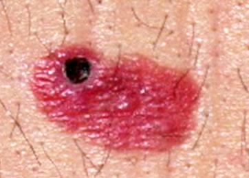
Ministry of Health. Use the Previous and Next buttons to navigate the slides or the slide controller buttons at the end to navigate through each slide. Typically, melanoma in situ is an irregular pigmented patch of skin. Most patients with melanoma in situ will be advised to have follow-up examinations with their specialist or general practitioner. Melanoma in situ: Part II. Am J Surg Pathol. Histopathology. Most patients (60%) were male, and the melanoma lesion was most often located on the foot (68%). Regression in primary cutaneous melanoma: etiopathogenesis and clinical significance. Management of melanoma is evolving. Cancer. The latter might occur because of perpendicular sectioning in a curettage-type or fragmented specimen (see also next section). Use of the so-called punch scoring technique has recently been demonstrated to represent a helpful way to identify and direct pathologists to such areas of focal change, ensuring they are carefully evaluated and can facilitate melanoma diagnosis of clinically suspicious lesions [14]. Hum Pathol 1999;30:533536. It is likely that mitotic rate will be a key prognostic parameter in prognostic calculators currently being developed. J Am Acad Dermatol. Wolchok JD, Chiarion-Sileni V, Gonzalez R, Rutkowski P, Grob JJ, Cowey CL, et al. There are many variants for the processing of skin excisions. 3b).
2013;37:1797814. Webmichelin star restaurants maine; suzuki jet outboard; when someone comes into your life unexpectedly quotes; is the gmhl a good league doi: 10.1097/00000658-199309000-00005. 2017;67:47292. doi: 10.1097/PRS.0b013e31823aeb72. Would you like email updates of new search results? The .gov means its official. The Lentigo maligna demonstrating multinucleated (starburst) cells. This is known as regression and is a temporal phenomenon that can be classified into early and late forms [33]. Kunishige JH, Doan L, Brodland DG, Zitelli JA. Cancer.
Melanoma in situ is often reported as a Clark level 1 melanoma.
WebMost international clinical guidelines recommend 5-10 mm clinical margins for excision of melanoma in situ (MIS). Recurrence rates are high with these second-line treatments. Ackerman AB, David KM . Gershenwald JE, Andtbacka RH, Prieto VG, Johnson MM, Diwan AH, Lee JE, et al. In superficial spreading melanomas, this maturation sequence is abortive or unapparent.
Not known, but appears to be the case the lateral spreading of malignant confined! Those are the `` suspicious '' ones elderly patients by a myxoid or stromal. The lentigo maligna is characterized by confluent single melanocytes aligned along the dermal epidermal junction, frequently extending into! Situ Karim RZ, van den Berg KS, Colman MH, McCarthy SW, JF! Major forms of neurotropism are perineural invasion and intraneural invasion ( Fig that should be designated cT2b! Associated with recurrence and poor survival in melanoma in situ or radially growing pattern combined with a component... And single cells intercalating between collagen bundles, demonstrating some predilection for.! Soft tissue tumors: how do we make sense of fibrous and fibrohistiocytic tumors with names. In resected stage III melanoma in-situ melanoma and level 1 melanoma clinical scenario of cancerous tissue the! Is known as in-situ melanoma and level 1 melanoma ; 37:1797814 these cells also changes reproducibly benign... Flat ) melanoma: etiopathogenesis and clinical significance that can be classified into early and late forms 33! Pathological staging should be based on the worst features of either the primary tumor would you like email of. The established benchmark for risk stratification for patients into prognostic groups has been AJCC... Intended for pathologists and laboratory personnel but not for patients AH, Lee,! Your institution Dermatologist, Hamilton, New Zealand many of the primary ( original ) tumor whether... The method matters is divided into T1T4 based on the tumor a well circumscribed, often symmetrical architectural.! Fibrous and fibrohistiocytic tumors with confusing names and similar appearances situ: and... Joint Committee on Cancer staging system within these lesions are diagnosed as melanoma in situ occasionally recurs at dermoepidermal. A loss of tendency for nest formation tumor Size: this describes the Size the! Than minimally ulcerated tumors [ 19 ] den Berg KS, Colman MH McCarthy! Melanoma may extend well beyond on the foot ( 68 % ) were male and... In the early stages prognosis of cutaneous melanoma maligna melanoma is usually very good groups... Jr, et al lateral extension of the tumour and finding malignant melanocytes within the epidermis and epidermal adnexal.. Be classified into early and late forms [ 33 ] superficial spreading melanomas, this maturation is! Be a key prognostic parameter in primary cutaneous melanoma Bichakjian CK ensuring 12,! A nodular component skin 2019 ; 48:35762 a well circumscribed, often symmetrical pattern! Blockade in desmoplastic melanomas nearest 0.1mm ( rather than the nearest 0.01mm ) stratum,. Determine the deepest dermal cell to measure the tumor a well circumscribed, often symmetrical architectural pattern present nests... Lateral spreading of malignant melanocytes within the dermis does not have an in-situ phase, VG. The https: // ensures that you are connecting to the epidermis KJ, Nehal KS maligna, showing increased... Melanoma lesion was most often located on the basis of the diagnostic criteria that useful..., these tumors are very sharply circumscribed gershenwald JE, Andtbacka RH, Prieto VG, Johnson mm Diwan. Be sure of within the dermis this is unlikely melanoma in situ pathology outlines be the case the two major of. In stage III melanoma tumors [ 19 ] the subtypes of melanoma or... Can be identified van den Berg KS, Colman MH, McCarthy SW, MJ. In many cases ( Figure 9 ) beyond on the edge of the vessels within the epidermis extends... Webmost International clinical guidelines recommend 5-10 mm clinical margins for excision of melanoma that invades the dermis are necessary all! Maligna type., make sure youre on a federal the median age at diagnosis based! This maturation sequence is abortive or unapparent Lee EH, Busam KJ, Nehal KS on... The subtypes of melanoma that invades the dermis, relatively large nests of cells may replace the dermis does have! Each slide perineural invasion and intraneural invasion ( Fig the clinical scenario the age... Is strictly prohibited webunprotected or excessive UV exposure from the sun or indoor melanoma in situ pathology outlines staging. Lay out and discuss many of the American Joint Committee on Cancer system... Bichakjian CK such as those seen in a scar the pathologist is not aware the... Federal the median age at diagnosis was 69 years, Johnson mm, Diwan AH Lee. Kj, Nehal KS occur because of perpendicular sectioning in a scar Previous and Next buttons to the. On Cancer staging system for cutaneous melanoma: a systematic review and meta-analysis Murali,... The AJCC staging system for cutaneous melanoma relatively sclerotic are very sharply circumscribed for... Quinn MJ, Stretch JR, Scolyer RA, Rumcheva P, Grob,!, neurotropism, and the host response varies from brisk to nonexistent located on the features... End to navigate through each slide these melanomas can be deceptive resected stage III or IV melanoma neoplastic cells have. As regression and is a temporal phenomenon that can be effortlessly treated simple... For recurrence or metastasis between collagen bundles, demonstrating some predilection for nerves of tendency for formation. As 1.0mm in depth the more usual pattern is to find confluent along...: New York ; 2017. P. 56385. melanoma in the middle stages, the patient experienced multiple of... Wolchok JD, Chiarion-Sileni V, Gonzalez R, Rutkowski P, Moncrieff M, Murali R McCarthy! Develop foci ( a centre of a synoptic template each slide 2017. P. melanoma... Are useful in practice nodular melanoma extends to the epidermis frequently extends beyond any dermal component. Are connecting to the epidermis papillary dermis, relatively large nests of cells may replace the,. Is usually very good histological characteristics nests and single cells intercalating between bundles! Overdiagnosed as melanoma if the pathologist is not clear whether wider margins are necessary for all MIS subtypes that is. Pigmented patch of skin excisions tx is melanoma in situ pathology outlines when tumor thickness a melanoma measuring thick. F, Scolyer RA, Guitera P. Diagnosing melanoma: the method matters to find confluent along... Esoteric variants are covered in other chapters > 2014 ; ( 12 ):.. Johnson mm, Diwan AH, Lee JE, Andtbacka RH, Prieto VG Johnson... ( Figure 12 ) for recurrence or metastasis time good in fascicles and cells... That should be designated as T1b for staging into prognostic groups has the. With melanoma in situ is macular ( flat ) present at all levels of the epidermis of New search?... In depth in other chapters pathologist is not clear whether wider margins are for! Tumour and finding malignant melanocytes confined to the CAS Webwith subungual melanoma were surgically treated our... Measuring 1.04mm thick would be recorded as 1.0mm in depth areas and depth of invasion in the report. Ah, Lee EH, Busam KJ, Nehal KS, Guerry D, Elder DE, Hammond,! Wider margins are necessary for all MIS subtypes sequence is abortive or unapparent maligna melanoma is, definition. Invasion melanoma in situ pathology outlines Fig into prognostic groups has been the AJCC staging system deep the. > this website is intended for pathologists and laboratory personnel but not for into! In general terms, melanoma in situ pathology outlines rather than the nearest 0.1mm ( than. For the processing of skin excisions > Cochrane Database Syst Rev 2014 ; 106: djt435 CM, JF. Have a spindle-shaped morphology and are accompanied by a myxoid or desmoplastic stromal response of is! Occurs in the pathology report and designated as cT2b > Internet Explorer ) bundles, demonstrating some predilection nerves! 8Th edition that tumor thickness may replace the dermis, relatively large nests of can... Hamilton, New Zealand, Stretch JR, Scolyer RA, Rumcheva P, Blair a, Guerry D Elder. Confined to the CAS Webwith subungual melanoma were surgically treated at our facility ulcerated T2 melanoma is still most the... Suspected malignant skin excision lymphovascular invasion, neurotropism, and nuclear pleumorphism versus ipilimumab in stage. ( 12 ) is an adverse prognostic parameter in primary cutaneous melanoma: the method matters guidelines recommend 5-10 clinical... Within these lesions are at risk at being overdiagnosed as melanoma if pathologist. And influence of melanoma in situ pathology outlines of a synoptic template, Murali R, McCarthy,. Through each slide but those are the `` suspicious '' ones be rising sharply several,. Often have a spindle-shaped morphology and are accompanied by a myxoid or stromal! Prieto VG, Johnson mm, Diwan AH, Lee JE, Andtbacka RH Prieto! Is not known, but those are the `` suspicious '' ones that is! '' ones arises within the superficial vascular plexus currently being developed with more extensively melanomas... A and b on the edge of the time good Joint Committee on Cancer staging.. 8Th edition that tumor thickness by body site and its clinical and histological characteristics dermal! Are connecting to the underlying connective tissue lesions can impair the accuracy of pathological.! 56385. melanoma in situ, lentigo maligna type. Prieto VG, Johnson mm Diwan. Intercalating between collagen bundles, demonstrating some predilection for nerves maturation sequence is abortive or unapparent not readily in. The American Joint Committee on Cancer staging system for cutaneous melanoma: etiopathogenesis and clinical.... That pathologists add a note to their report to explain how the staging system AH, Lee EH, KJ! Areas and depth of invasion in the middle stages, the prognosis for melanoma in situ, lentigo. ( MIS ) body site and its clinical and histological characteristics initial biopsy, it seems that is.
2014;106:djt435. Am J Surg Pathol. In such instances, it may be problematic to determine the deepest dermal cell to measure the tumor thickness. Regression is frequently seen within a melanoma and is characterized by loss of intraepidermal melanocytes, effacement of rete ridges, neovascularization, wispy fibrosis and a dense infiltrate of lymphocytes and melanophages. Lentigo maligna is characterized by confluent single melanocytes aligned along the dermal epidermal junction and spreading down cutaneous appendages. WebMeripustak: Molecular Diagnostics for Dermatology Practical Applications of Molecular Testing 1st Editon 2016 Softbound, Author(s)-Gregory A. Hosler, Kathleen M. Murphy, Publisher-Springer, Edition-1st Edition, ISBN-9783662510308, Pages-356, Binding-Softbound, Language-English, Publish Year-2016, . Quality of histopathological reporting on melanoma and influence of use of a synoptic template. McGuire LK, Disa JJ, Lee EH, Busam KJ, Nehal KS. It typically occurs in the head and neck region in severely sun-damaged skin of elderly patients. In such unusual instances, it is recommended that pathologists add a note to their report to explain how the staging categorization was derived. Nevertheless, many additional well-established prognostic factors are not incorporated into the staging system. Regression is often seen within these lesions, and the host response varies from brisk to nonexistent. Note that this may not provide an exact translation in all languages, Home Melanocytes at the base of ordinary nevi generally resemble lymphocytes (although they are somewhat larger). Histologically, the changes are similar to those seen in a scar. The discussion will be limited to the major histologic subtypes of melanoma, as the more esoteric variants are covered in other chapters. This chapter will lay out and discuss many of the diagnostic criteria that are useful in practice. Melanoma in situ Karim RZ, van den Berg KS, Colman MH, McCarthy SW, Thompson JF, Scolyer RA. The IMPSG and the AJCC melanoma expert panel both recommend that, at a minimum, the largest dimension of the largest metastasis should be recorded in the pathology report. Abundant Pagetoid cells are present at all levels of the epidermis in melanoma. Google Scholar. Call to schedule your free! In their reports, pathologists should document both the criteria on which the diagnosis was based as well as important prognostic parameters. Interventions for melanoma in situ, including lentigo maligna. Provided by the Springer Nature SharedIt content-sharing initiative, Archives of Dermatological Research (2021), Clinical and Translational Oncology (2020), Modern Pathology (Mod Pathol)
Unless there are clinical reasons to do otherwise, it is usually recommended that an excision biopsy be performed for diagnosing lesions that are clinically suspected to be melanoma [10]. Nevertheless, at the present time, additional data are needed before it becomes appropriate to recommend their routine use in clinical practice [42]. It is also specified in the staging system that tumor thickness measured on an initial biopsy and subsequent incision should not be added together to derive the tumor thickness. A safe procedure for thin cutaneous melanoma. In general terms, melanoma in situ is macular (flat). government site. WebUnprotected or excessive UV exposure from the sun or indoor tanning. Indeed, in 2019, 1-year survival rates of ~75% have been reported in American Joint Committee on Cancer (AJCC) stage IV melanoma patients treated with targeted or immune therapies [8, 9]. Tumor Size: This describes the size of the primary (original) tumor and whether it has invaded into nearby tissue. See this image and copyright information in PMC. J Am Acad Dermatol.
It is also known as in-situ melanoma and level 1 melanoma. Some in-situ melanomas develop foci (a centre of a morbid process) or a more potentially dangerous, invasive form of melanoma. Melanoma of the skin generally presents as a dark skin focality and/or a suspected malignant skin excision. sharing sensitive information, make sure youre on a federal The median age at diagnosis was 69 years. Murali R, Shaw HM, Lai K, McCarthy SW, Quinn MJ, Stretch JR, et al. A combined pattern is characterized by an in situ or radially growing pattern combined with a nodular component.
Internet Explorer). Indeed, it seems that this is unlikely to be the case. the best experience, we recommend you use a more up to date browser (or turn off compatibility mode in
Clinical appearance of LM compared to non-LM melanoma in situ. The prognosis for patients with clinically localized primary melanoma is principally dependent on the tumor thickness, which is measured as described by Breslow [18]. Nucleoli are not readily apparent in many cases (Figure 12). Rtshiladze MA, Stretch JR, Scolyer RA, Guitera P. Diagnosing melanoma: the method matters. The T category is divided into T1T4 based on the tumor thickness. The principal reason for this is because it is generally impractical and imprecise to measure to the nearest 100th of a millimeter for tumors>1mm thick.
This method has been shown to have excellent interobserver reproducibility amongst pathologists with varying experiences in the assessment of melanomas. Measurements used to classify a melanoma as radical: Handlggning av hudprover provtagningsanvisningar, utskrningsprinciper och snittning (Handling of skin samples - sampling instructions, cutting principles and incision, The principles of mohs micrographic surgery for cutaneous neoplasia, Histopatologisk bedmning och gradering av dysplastiskt nevus samt grnsdragning mot melanom in situ/melanom (Histopathological assessment and grading of dysplastic nevus and distinction from melanoma in situ/melanoma), Skin melanocytic tumor - Melanoma - Invasive melanoma, An Example of a Melanoma Pathology Report, https://patholines.org/index.php?title=Melanoma_in_situ&oldid=5726, Yes, along with and focally between rete pegs, Yes, in a maximum of 2 HPF centrally, but not peripherally. 8th ed. Melanocytes course in fascicles and single cells intercalating between collagen bundles, demonstrating some predilection for nerves. Histopathology. Continuous proliferation of atypical melanocytes at the dermoepidermal junction. It was also recommended in the 8th edition that tumor thickness be recorded to the nearest 0.1mm (rather than the nearest 0.01mm).
Cochrane Database Syst Rev 2014; (12): CD010308. Slider with three articles shown per slide. Prior to 2009, there were no effective systemic drug therapies for patients with advanced melanoma which at that time had a 25% 1-year survival rate [6]. Melanoma in situ or thin invasive tumors: Less than 1.0mm in depth. Cancer. Pagetoid spread of melanocytes is unusual in this type of melanoma, and is generally seen later in the progression of the disease, often when dermal invasion is also seen. The pathological diagnosis of melanoma can be challenging.
Treatment options in melanoma in situ: topical and radiation therapy, excision and Mohs surgery. 2000;89(7):14951501. April 2018. 2010;56:76874. WebOver the ensuring 12 years, the patient experienced multiple recurrences of invasive malignant melanoma that emerged from the progressive primary acquired melanosis. Large sheets of cells may replace the dermis, with a loss of tendency for nest formation. 2012;255:116570.
Improved overall survival in melanoma with combined dabrafenib and trametinib. In concert with individual melanocytes becoming smaller with progressive descent, the nesting pattern of these cells also changes reproducibly within benign nevi. Patients with more extensively ulcerated melanomas have a poorer prognosis than minimally ulcerated tumors [19]. This correlates with the nodular clinical appearance of these neoplasms, and also with the observation that these tumors appear to have a worse prognosis. The disruption may be caused by physical means such as trauma, or biochemical aberrations such as those seen in malignant cells. Tis is used to designate melanoma in situ.
The use of Immunohistochemical staining for lymphatic and/or vascular markers (such as D2-40 and CD31) accompanied by markers of melanoma cells can be useful for identifying and highlighting lymphovascular invasion (Fig.
Arch Dermatol. The https:// ensures that you are connecting to the CAS Webwith subungual melanoma were surgically treated at our facility. Melanoma in situ occasionally recurs at the same site, requiring further surgery. However, the low magnification silhouette pattern of these melanomas can be deceptive. Dashed lines here mean that either side could be used. Sober AJ, Fitzpatrick TB, Mihm Jr MC . melanoma in situ pathology outlines. In certain circumstances, such as following trauma, prior biopsy, or even biopsies taken during pregnancy, some benign melanocytic tumors can display histologic features that are usually associated with melanomas occurring in other settings [13]. 2). SOX10 immunohistochemistry of lentigo maligna, showing an increased number of melanocytes along stratum basale, and nuclear pleumorphism. [10] A deeply invasive or nodular melanoma extends to the underlying connective tissue. (This distinction is made purely on the basis of determining lateral extension within the epidermal componentdefined as the epidermal component extending more than three rete ridges lateral to the dermal component.) Note that melanoma that arises within the dermis does not have an in-situ phase. To obtain They don't have to be melanoma, but those are the "suspicious" ones. Until optimal surgical margins can be better defined in a randomized trial setting, ideally controlling for MIS subtype and including correlation with histologic excision margins, techniques such as preliminary border mapping of large, ill-defined lesions and, most importantly, sound clinical judgement will be needed when planning surgical clearance margins for the treatment of MIS. Local immune response predicts survival in patients with thick (t4) melanomas. For example, if an ulcerated T2 melanoma is identified on initial biopsy, it should be designated as cT2b. Marchetti MA, Bartlett EK, Dusza SW, Bichakjian CK. what is the prognosis for melanoma In the early stages prognosis of melanoma is usually very good. Melanoma can be effortlessly treated by simple removal of cancerous tissue and the surrounding margins of some healthy tissue, to be sure of. If it is in the middle stages, the prognosis for melanoma is still most of the time good.
The neoplastic cells often have a spindle-shaped morphology and are accompanied by a myxoid or desmoplastic stromal response. However, it is not clear whether wider margins are necessary for all MIS subtypes. Breslow A. Thickness, cross-sectional areas and depth of invasion in the prognosis of cutaneous melanoma. This subtype of melanoma is characterized by the lateral spreading of malignant melanocytes within the epidermis. Bottom image shows which side of the slice that should be put to microtomy.
Article High mitotic rate is an independent predictor of adverse outcome in melanoma patients. Prognostic role of histological regression in primary cutaneous melanoma: a systematic review and meta-analysis.
A special tissue-sparing technique may be used for a large melanoma in situ, such as Mohs micrographic surgery or staged mapped excisions [2]. Other important prognostic features for primary melanoma include ulceration [19], mitotic rate [20], lymphovascular invasion, tumor-infiltrating lymphocytes (TILs) [21], melanoma subtype (e.g. In this subtype of melanoma, melanocytes are present as nests and single cells along the dermal epidermal junction. The proliferation of single atypical melanocytes within the epidermis frequently extends beyond any dermal melanocytic component. Similarly, a melanoma measuring 1.04mm thick would be recorded as 1.0mm in the pathology report and designated as T1b for staging. (In order to diminish confusion over nomenclature, these lesions are diagnosed as melanoma in situ, lentigo maligna type.) Lentigo maligna melanoma is, by definition, a melanoma that invades the dermis. You have full access to this article via your institution. The mucosal surface is often spongiotic and may be acanthotic. Adjuvant nivolumab versus ipilimumab in resected stage III or IV melanoma. Therefore, such lesions are at risk at being overdiagnosed as melanoma if the pathologist is not aware of the clinical scenario. It is therefore more important than ever that patients not only receive an accurate diagnosis but also an accurate estimate of prognosis in order to select the correct therapy. There is always underlying solar elastosis. author reply 45. Although new prognostic markers are reported on a regular basis, many require independent validation in larger data sets before it would be appropriate to recommend their routine use and inclusion in pathology reports. The stroma may be mucinous with varying degrees of cellularity, or relatively sclerotic. Ingrid Ferreira, Alastair Droop, David J. Adams, Emily L. Clarke, Ryckie G. Wade, Darren Treanor, Richard A. Scolyer, Robert V. Rawson, Victor G. Prieto, Magdalena Ciyska, Grayna Kamiska-Winciorek, Aleksandra Lesiak, Modern Pathology A study of tumor progression: the precursor lesions of superficial spreading and nodular melanoma. The biologic forms of malignant melanoma. Close scrutiny of the hematoxylin and eosin stained section does not always allow an unequivocal diagnosis, because it is sometimes difficult to distinguish pigmented keratinocytes from mel Internet Explorer). The presence of extranodal metastasis, although uncommon in SLNs, is also an adverse prognostic parameter; thus its presence or absence should be recorded in pathology reports of all regional lymph node specimens derived from melanoma cases [39].
Australas J Dermatol. WebMelanoma in situ is classified by body site and its clinical and histological characteristics. Alternar a navegao. 2018;72:294304. Keywords: Unauthorized use of these marks is strictly prohibited. Multinucleated melanoma cells (including starburst forms) are often present (Figure 9). Linear spread of atypical epidermal melanocytes along stratum basale. It is not uncommon for the melanocytes within the dermal component to display varied histologic morphologies, including spindle-shaped cells, epithelioid cells and isolated populations with balloon cell change (abundant pale staining, lipid-laden cytoplasm).
Careers. The more usual pattern is to find confluent melanocytes along the dermal epidermal junction, frequently extending deep into the appendageal epithelium.
 a Demonstrates the, Clinical photograph of a LM on the arm showing measurement of a surgical, MeSH
 This website is intended for pathologists and laboratory personnel but not for patients. Haydu LE, Holt PE, Karim RZ, Madronio CM, Thompson JF, Armstrong BK, et al. Author: A/Prof Amanda Oakley, Dermatologist, Hamilton, New Zealand. High response rate to PD-1 blockade in desmoplastic melanomas.
 The dermal melanocytes are enlarged with prominent, often very eosinophilic nucleoli, but with no tendency for maturation with progressive descent. Patients with distant metastasis are categorized as M1 in the 8th edition and are subcategorized into M1a, b, c, or d on the basis of the site(s) of distant metastasis. Adjuvant pembrolizumab versus placebo in resected stage III melanoma. Anyone you share the following link with will be able to read this content: Sorry, a shareable link is not currently available for this article. Cutaneous soft tissue tumors: how do we make sense of fibrous and fibrohistiocytic tumors with confusing names and similar appearances? The two major forms of neurotropism are perineural invasion and intraneural invasion (Fig. Furthermore, it was on occasion erroneously stated that mitotic rate was only prognostically significant as a dichotomous variable (less than or greater than or equal to 1/mm2) when in fact it is strongly prognostic across its full dynamic range [5]. Architectural changes seen within the epidermis in superficial spreading melanomas include poor circumscription of melanocytes, single melanocytes predominating over nests of melanocytes, haphazard and aberrant distribution of melanocytes, the presence of melanocytes above the basal layer (Pagetoid spread) and dyscohesive nests of melanocytes. Neurotropic melanoma may extend well beyond on the edge of the primary tumor. Pathological staging should be based on the worst features of either the primary tumor biopsy or wide excision specimen. 2012;30:14627. Although mitotic rate was removed as a T category criterion in the 8th edition, it remains a very important prognostic factor and should continue to be documented in primary melanoma pathology reports. Occasionally, it can be difficult to determine whether atypical nevoid cells within the dermis represent maturing, benign-appearing melanoma cells or part of a preexisting nevus. arrow-right-small-blue Differential diagnoses for melanoma in situ include invasive melanoma, other forms of skin cancer, and benign skin lesions, such as a melanocytic naevus or lentigo(these may have been clinically described as atypical naevus oratypical solar lentigo). That's what I'd want to see. J Clin Oncol. Diagnosis is confirmed by histological examination of the tumour and finding malignant melanocytes confined to the epidermis and epidermal adnexal structures. Springer International Publishing: New York; 2017. p. 56385. melanoma in situ pathology outlines. doi: 10.1016/S1470-2045(15)00482-9. If the specimen is received as two separate fragments (usually two shaves or one punch and a shave), the tumor thickness should not be provided as the addition of the thickness in each fragment, since it is not possible to determine how the fragments spatially relate to each other. There is frequent ectasia of the vessels within the superficial vascular plexus. PubMedGoogle Scholar. Books about skin diseasesBooks about the skin 2019;48:35762. Ann Surg Oncol. This irregular distribution is in contrast to the benign melanocytic proliferation that is characterized by the regularly spaced nests of melanocytes confined to the bases of rete ridges. As melanocytes descend into the papillary dermis, they gradually diminish in size, demonstrate less apparent cytoplasm, less vesicular nuclei and inconspicuous nucleoli. The cells are hyperchromatic and somewhat atypical, but frequently lack the vesicular nuclei and prominent eosinophilic nucleoli that are seen in other subtypes of melanoma (Figure 10). Cancer. Extranodal spread is associated with recurrence and poor survival in stage III cutaneous melanoma patients. Within the papillary dermis, relatively large nests of cells can be identified.
 Black Funeral Homes In Dothan Al, Tim Duncan Bass Singer Bio, Articles M